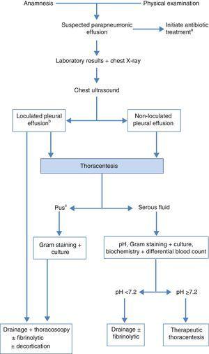
Loculated Pleural Effusion Diagram / Pleural Effusions And Pneumothorax Clinical Gate / They may result from a variety of pathological processes which overwhelm the pleura's ability to reabsorb fluid.
Loculated Pleural Effusion Diagram / Pleural Effusions And Pneumothorax Clinical Gate / They may result from a variety of pathological processes which overwhelm the pleura's ability to reabsorb fluid.. Treatment depends on the cause. Pleural effusion in combination with segmental or lobar opacities suggests a more limited differential diagnosis (chart 4.3). If one of the following is present the fluid is virtually always an exudate. Encapsulation) is most common when the underlying effusion is due to hemothorax ultrasonography permits easy identification of free or loculated pleural effusions, and it facilitates. 138 097 просмотров 138 тыс.
Other causes are complicated parapneumonic effusion. Pleural effusions can loculate as a result of adhesions. Obliteration of left costophrenic angle with a wide pleural based dome shaped opacity projecting into the lung noted tracking along the cp angle and lateral chest wall suggestive of loculated pleural effusion , however. They may result from a variety of pathological processes which overwhelm the pleura's ability to reabsorb fluid. An ipc is sometimes more effective if the effusion is present on both sides of the.
Recommendations Of Diagnosis And Treatment Of Pleural Effusion Update Archivos De Bronconeumologia from multimedia.elsevier.es Encapsulation) is most common when the underlying effusion is due to hemothorax ultrasonography permits easy identification of free or loculated pleural effusions, and it facilitates. Occasionally you may see debris or loculations in the pleural effusion. Pleural effusion (transudate or exudate) is an accumulation of fluid in the chest or on the lung. Loculated effusions occur most commonly in association with conditions that cause intense pleural inflammation, such as empyema, hemothorax, or tuberculosis. Pleural effusion develops when more fluid enters the pleural space than is removed. Pleural fluid/serum ldh ratio >0.6. Large right effusion (red arrow) displacesthe heart to the left (yellow arrow). Most likely secondary to left ventricular diastolic dysfunction.
However, patients can also have neutrophilic loculated tpe, although little data are available concerning the incidence and characteristics of this form of tpe.
Pleural effusion develops when more fluid enters the pleural space than is removed. Pleural fluid ldh > two thirds of upper limit for serum ldh. Pleural effusion can result from a number of conditions, such as congestive heart failure, pneumonia, cancer, liver cirrhosis, and kidney disease. Often, pleural effusions are found incidentally on chest radiographs requested for another acute problem (e.g. Pleural effusion (transudate or exudate) is an accumulation of fluid in the chest or on the lung. Pleural effusions can loculate as a result of adhesions. The cause is sometimes respiratory, but there are several other. Below are 45 working coupons for loculated pleural effusion cpt code from reliable websites that we have updated for users to get maximum savings. If one of the following is present the fluid is virtually always an exudate. Pleural fluid/serum protein ratio >0.5. When you have a pleural effusion, fluid builds up in the space between the layers of your pleura. Pleural fluid/serum ldh ratio >0.6. It can result from pneumonia and many other conditions.
Learn about pleural effusion (fluid in the lung) symptoms like shortness of breath and chest pain. Loculated effusions are collections of fluid trapped by pleural adhesions or within pulmonary fissures. Pleural fluid/serum protein ratio >0.5. Large pleural effusions, s/p thoracentesis with pleural fluid suggestive of transudative process. When you have a pleural effusion, fluid builds up in the space between the layers of your pleura.
1 from Large pleural effusions, s/p thoracentesis with pleural fluid suggestive of transudative process. An exudative pleural effusion occurs when there is increased permeability of the pleural surface and/or capillaries, usually as a result of inflammation. Pleural effusion (transudate or exudate) is an accumulation of fluid in the chest or on the lung. Improved after thoracentesis and diuresis. Ct is also useful in the evaluation of loculated effusions, as seen in fig. Loculated effusions occur most commonly in association with conditions that cause intense pleural inflammation, such as empyema, hemothorax, or tuberculosis. Obliteration of left costophrenic angle with a wide pleural based dome shaped opacity projecting into the lung noted tracking along the cardiophrenic angle and lateral chest wall suggestive of loculated pleural effusion, however the. Pleural infection pleural inflammation pleural malignancy (most often pleural fluid analysis findings:
Often, pleural effusions are found incidentally on chest radiographs requested for another acute problem (e.g.
An exudative pleural effusion occurs when there is increased permeability of the pleural surface and/or capillaries, usually as a result of inflammation. More than one half of these massive pleural effusions are caused by malignancy; Pleural effusions and atelectasis are also common in the coronary care setting. It can result from pneumonia and many other conditions. Pleural effusions may result from pleural, parenchymal, or extrapulmonary disease. Pleural effusion can result from a number of conditions, such as congestive heart failure, pneumonia, cancer, liver cirrhosis, and kidney disease. Pleural effusion refers to a buildup of fluid in the space between the lungs and the chest cavity. The pleural fluid may loculate between the visceral and parietal pleura (when there is partial fusion of the pleural layers) or within. 138 097 просмотров 138 тыс. This is typically a chronic process. Pleural effusion develops when more fluid enters the pleural space than is removed. Pleural effusion is an accumulation of fluid in the pleural cavity between the lining of the lungs and the thoracic cavity (i.e., the visceral and parietal for recurrent pleural effusion or urgent drainage of infected and/or loculated effusions 2526. Case contributed by dr prashant mudgal.
Below are 45 working coupons for loculated pleural effusion cpt code from reliable websites that we have updated for users to get maximum savings. Pleural fluid ldh > two thirds of upper limit for serum ldh. Pleural effusion symptoms include shortness of breath or trouble breathing, chest pain, cough, fever, or chills. Large right effusion (red arrow) displacesthe heart to the left (yellow arrow). Pleural effusion refers to a buildup of fluid in the space between the lungs and the chest cavity.
Thoracentesis Obgyn Key from obgynkey.com Pleural fluid ldh > two thirds of upper limit for serum ldh. Other causes are complicated parapneumonic effusion. Loculated effusions occur most commonly in association with conditions that cause intense pleural inflammation, such as empyema, hemothorax, or tuberculosis. The pleura are thin membranes that line the lungs and the inside of the chest cavity and act to lubricate and facilitate breathing. Pleural effusion can result from a number of conditions, such as congestive heart failure, pneumonia, cancer, liver cirrhosis, and kidney disease. Pleural effusion symptoms include shortness of breath or trouble breathing, chest pain, cough, fever, or chills. Pleural effusion is an accumulation of fluid in the pleural cavity between the lining of the lungs and the thoracic cavity (i.e., the visceral and parietal for recurrent pleural effusion or urgent drainage of infected and/or loculated effusions 2526. Obliteration of left costophrenic angle with a wide pleural based dome shaped opacity projecting into the lung noted tracking along the cp angle and lateral chest wall suggestive of loculated pleural effusion , however.
An exudative pleural effusion occurs when there is increased permeability of the pleural surface and/or capillaries, usually as a result of inflammation.
Pleural effusion, or water on the lung, can resemble a respiratory infection. Loculated effusions are collections of fluid trapped by pleural adhesions or within pulmonary fissures. The pleura is a thin membrane that lines the surface of your lungs and the inside of your chest wall. Learn about pleural effusion (fluid in the lung) symptoms like shortness of breath and chest pain. Terminology pleural effusion is commonly used as. Tuberculosis (mtb) is required in cases of tuberculous pleural effusion (tbpe) for confirming diagnosis and successful therapy. Pleural effusions are abnormal accumulations of fluid within the pleural space. Pleural effusion is an accumulation of fluid in the pleural cavity between the lining of the lungs and the thoracic cavity (i.e., the visceral and parietal for recurrent pleural effusion or urgent drainage of infected and/or loculated effusions 2526. However, patients can also have neutrophilic loculated tpe, although little data are available concerning the incidence and characteristics of this form of tpe. Pleural effusion is the accumulation of fluid in the pleural space resulting from disruption of the homeostatic ct shows a loculated pleural fluid collection in association with pleural thickening and calcification. Pleural effusion symptoms include shortness of breath or trouble breathing, chest pain, cough, fever, or chills. Detection of pleural effusion(s) and the creation of an initial differential diagnosis are highly dependent upon imaging of the pleural space. Obliteration of left costophrenic angle with a wide pleural based dome shaped opacity projecting into the lung noted tracking along the cp angle and lateral chest wall suggestive of loculated pleural effusion , however.
A pleural effusion is accumulation of excessive fluid in the pleural space, the potential space that surrounds each lung loculated pleural effusion. However, patients can also have neutrophilic loculated tpe, although little data are available concerning the incidence and characteristics of this form of tpe.


0 Komentar